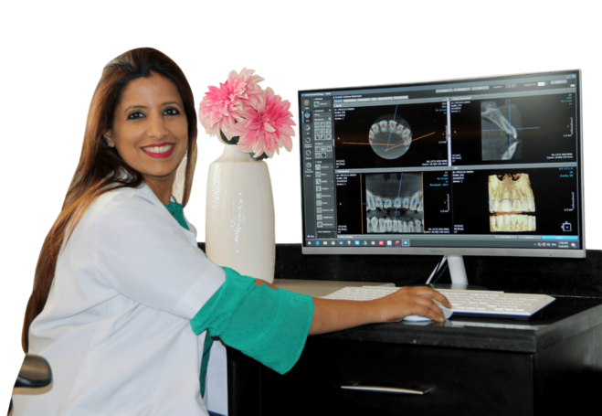
NEXT GENERATION ADVANCE DENTAL DIAGNOSTIC CENTRE FOR EXCELLENT TREATMENT PLAN
With your Dental visit, our team of Doctors make sure to fully understand your Dental problems in all the aspect keeping in mind to come up with perfect Diagnosis with 6 step Diagnostic workflow options in respect to the depth of problems always ensuring for optimal treatment plan
ADVANCE 6 DIAGNOSTIC WAYS FOR UNMATCHED DIAGNOSIS
RVG Single Tooth X-Ray Diagnosis
OPG Full Mouth X-Ray Diagnosis

CBCT Dental CT Scan 3D Diagnosis
VISUAL EXAMINATION
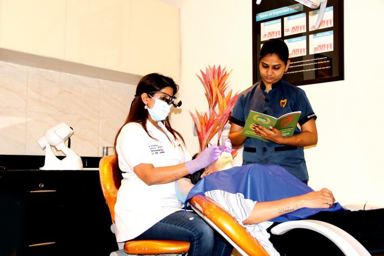
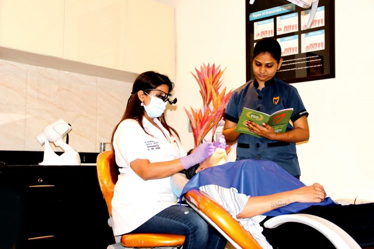
Complete overall Dental Examination is done inrespect to both the Hard and Soft Tissues in your Oral Cavity. All the Dental Findings are noted in a very professional & easy way so that you can understand them easily. We have well written performa that provides complete information of all the finding of the Hard Tissues and Soft Tissues after knowing complete Past Medical and Dental History.
Performa Consists :
-
Chief Complant
-
Soft Tissue Findings - Plaque, Calculus, Gingivitis
-
Hard Tissues Findings— Sensitivity, Decay, Filled Tooth, Impacted Tooth, Fractured Tooth, Fractured Filling, Missing Tooth, Mobility of Teeth, Pain on pressing the Tooth, RCT treated Tooth, Present Crown and Bridges, Present Implant Status.
SOPROCARE—DECAY & PLAQUE DIAGNOSTIC IMAGING
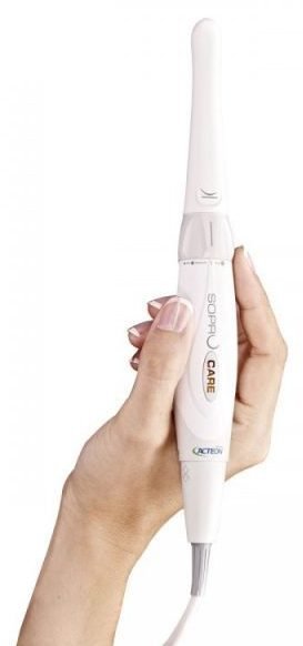

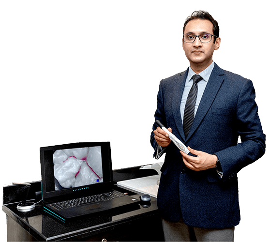
It’s a patented Imaging Device, that works on the basis of Auto-Flouroscence The device has got 3 modes
Cario Mode: To Diagnose the Dental Decay
Perio Mode: To Diagnose the Plaque and Gum Inflammation
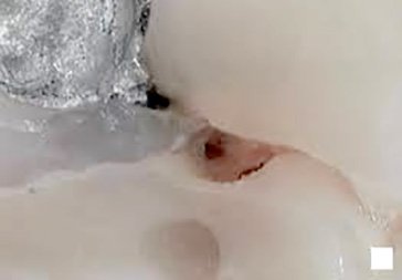
Daylight Mode: To Diagnose anatomical details that are invisible to naked eyes with 4 different Imaging Magnification.
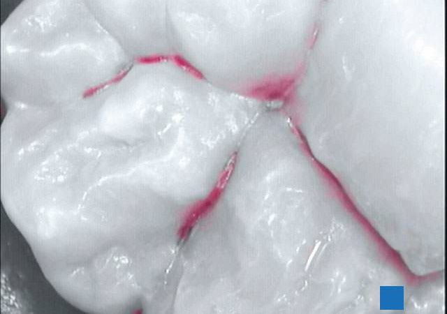
CARIO Mode
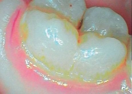
PERIO Mode
DAYLIGHT Mode
We made your Diagnosis Transparent
You can see your Decay & Plaque directly in front of TV Screen with Soprocare Patented Auto-Flouroscence Technology
Soprocare — Cario Mode
To Diagnose the Decay Digitally
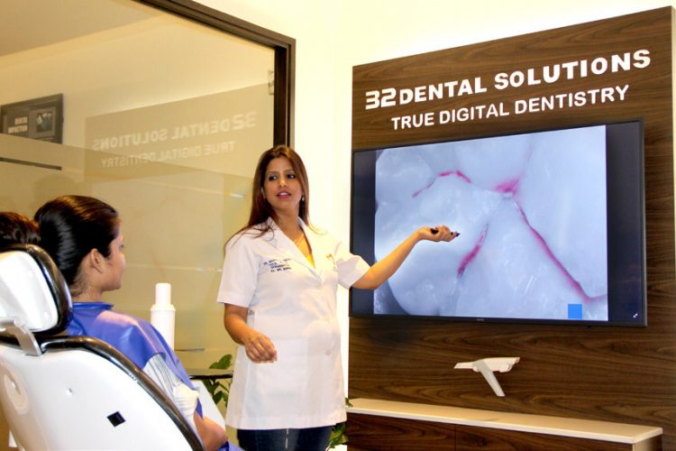
Confused between healthy Tooth or Decayed Tooth?? The answer is Soprocare Cario Mode—In Cario Mode, with Soprocare patented Auto-Flourosence Technology, Decays are shown as Bright Red Colour where as Healthy Tooth does not produce any colour with the Imaging
THERE ARE NO FALSE POSITIVE RESULTS, IF IT IS RED, IT’S A DECAY
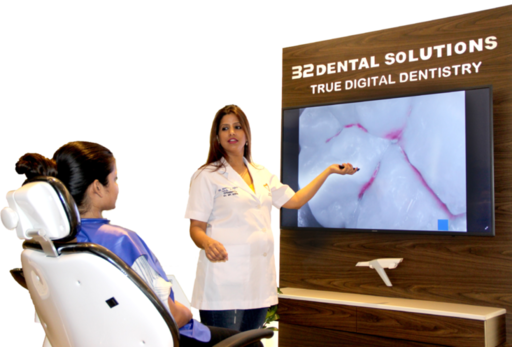
When our patients opt for Soprocare Decay Diagnosis, we scan the Tooth which had the possibility of Dental Decay using Soprocare Cario Mode An Image is Capurted & Appears on the TV Screen right in front of you showing the Tooth with or withour Red colour, which is then thoroughly explained by our team of Doctors as per the results to make an Accurate Treatment Plan
Soprocare — Perio Mode
To Diagnose the Plaque and Gum Inflammation
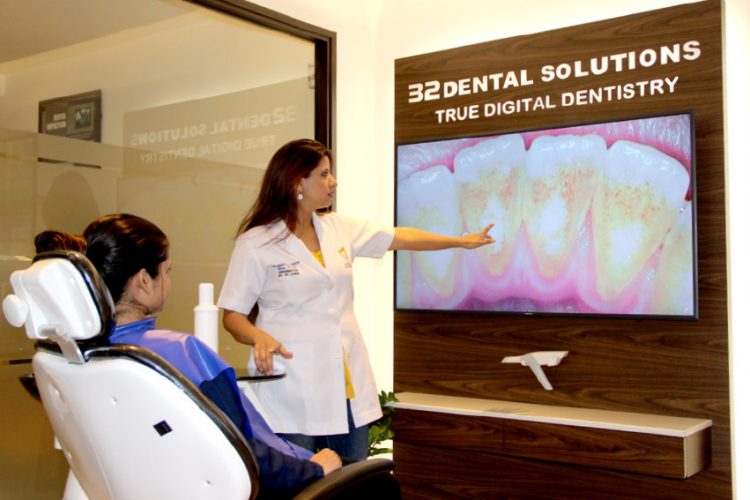
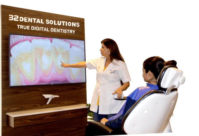
See your oral Hygiene status: with Soprocare Perio Mode. In Perio Mode our patients can see the Hidden Plaque and its effect on Gums as Inflammation which. The Dental Plaque is seen as Yellow Colour with Inflamed Gums are shown as Red Colour where as Healthy Tooth and Gums does not produce any colour
PLAQUE IS SEEN AS YELLOWISH RED COLOUR & INFLAMED GUMS AS DARK RED COLOUR
An image is capurted & appears on the TV screen right in front of you showing the Gums and Teeth. Gums if appeared Red are supposed to be Inflamed and Tooth surface if Yellowish-Red, indicates bacterial colony in the form of Plaque which is then thoroughly explained by our Team of Doctors as per the results to make an Accurate Treatment Plan
Soprocare — Day Light Mode
To See the Unseen
To Diagnose Anatomical details that are invisible to naked eyes with 4 different Imaging Magnification. When our patients opt for Soprocare Diagnosis, we scan the Tooth / Teeth in Soprocare Daylight Mode to see the unseen
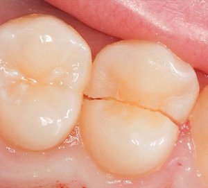
Cracked Tooth
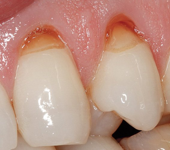
Abfraction Defects
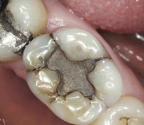
FractuRed Filling
In Daylight Mode the Soprocare Device can Diagnose and show any Abnormality with the Tooth such as Microcracks, FractuRed Filling, Abfraction Defects etc which are not visible with naked eyes with 4 different types on magnifiations
RVG – SINGLE TOOTH X-RAY DIAGNOSIS
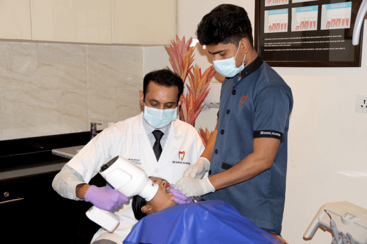
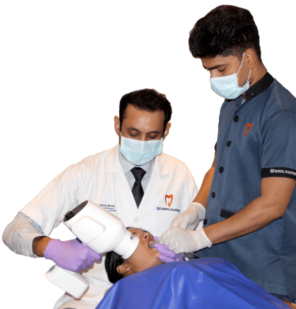
It is done to Diagnose problem related to single Tooth/single finding for knowing the extent of Decay, Mobility, Infection or other Problems related to Single Tooth
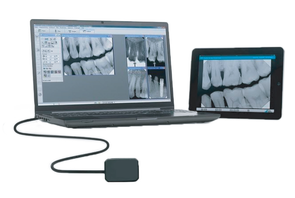
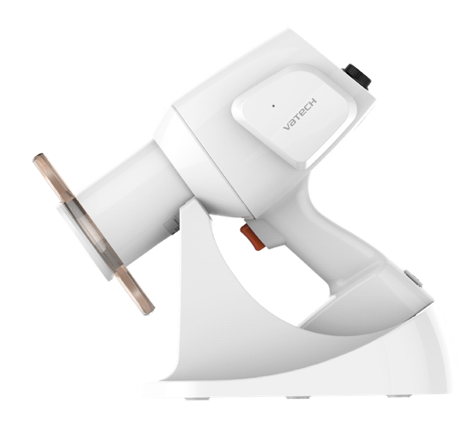
Vatech Digital RVG & X-Ray
We use state of the art- Vatech Digital Sensor (RVG) with Vatech X-Ray unit for Diagnostic Imaging of single Tooth.
Since it’s a digital X-Ray it used 1/10th of the radiation than conventional X-Ray and the X-Ray results comes instantly on to the computer software which is then thoroughly explained to our patients with an Accurate Treatment Plan.
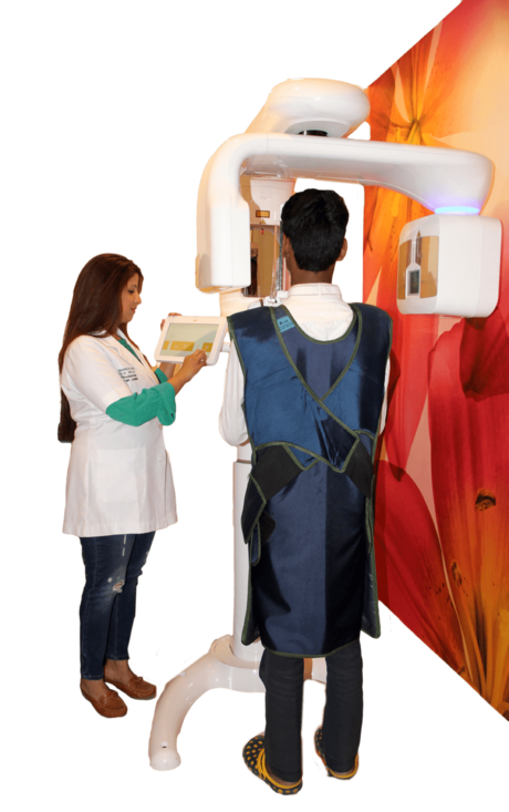
OPG—FULL MOUTH X-RAY DIAGNOSIS
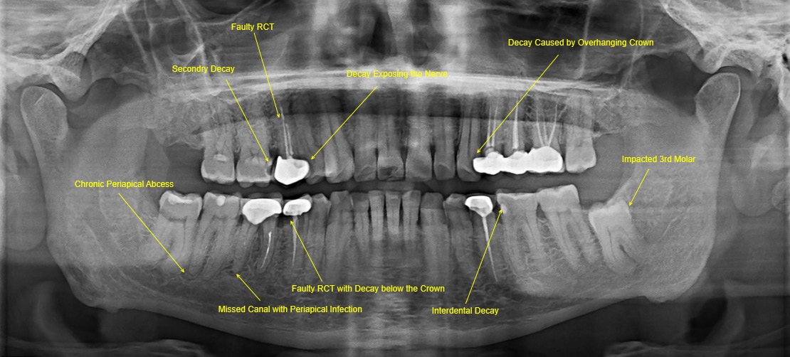
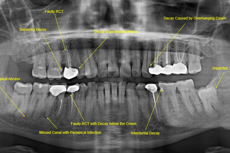
An OPG is a panoramic or wide view X-Ray of face, which displays all the Teeth of the upper and lower jaw on a single film. It demonstrates the number, position and growth of all the Teeth including those that have not yet surfaced or erupted.
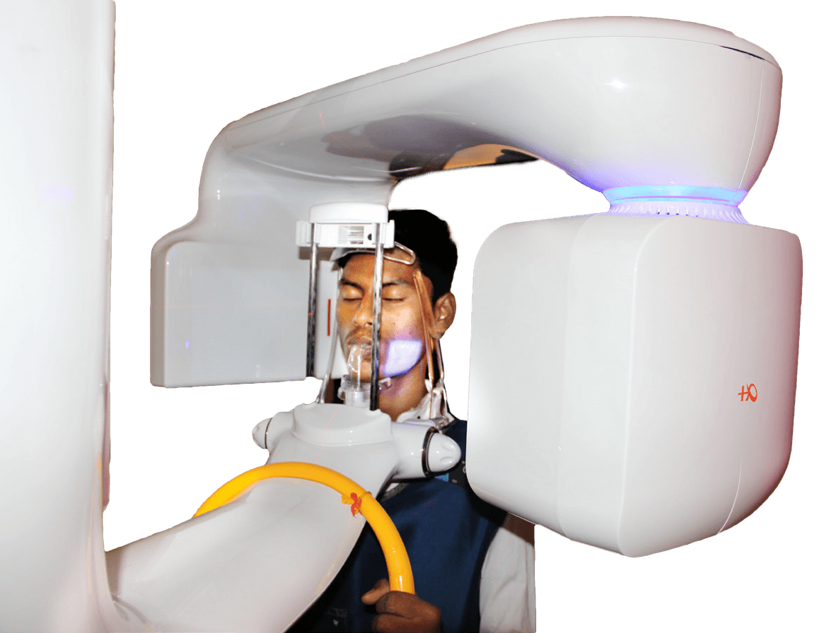
OPG is any time, a better choice as a Diagnostic Approach towards your Dental Problems
An OPG is a panoramic or wide view X-Ray of face, which displays all the Teeth of the upper and lower jaw on a single film. It demonstrates the number, position and growth of all the Teeth including those that have not yet surfaced or erupted.
